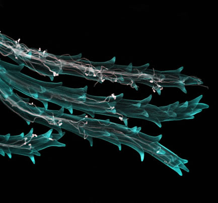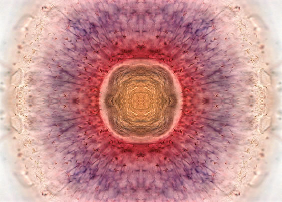
We’re used to taking pictures with our Nikon D5000s, our Olympus E-PL2s, and our Canon S95s. But there are some highly talented scientists out there making images with CT scanners, electron scanning microscopes, and confocal microscopes; astonishing pictures of moth wing scales, fish embryos, cells dividing, and plant stigmas. It’s not the sort of thing that you’ll find on Flickr, but you will find some of them – in fact a lot of them – at the Wellcome Collection.
Every year, the Wellcome Collection acquires thousands of images that document scientific exploration, development, and discovery. They’ve been doing this for a long time, but for the past 11 years, they’ve awarded prizes to the ‘most informative, striking and technically excellent’ images submitted to them that year.
The winners, all 21 of them, of the 11th Wellcome Image Awards have just been announced. It’s worth taking a look.
It’s also worth knowing that all of the images held by the Wellcome Collection are freely available for non-commerical personal and academic use. If you’d like to use images commercially, contact them to discuss fees.

Wheat infected with ergot fungus, Anna Gordon, National Institute of Agricultural Biology, AND Fernan Federici, University of Cambridge
If you can’t get to the Wellcome Collection to enjoy the exhibition, you can also peruse the gallery of winning images. You can also find out more about Wellcome Images in general.
Wellcome Image Awards exhibition runs until 10 July 2011 at the Wellcome Collection, 183 Euston Road, London, NW1 2BE.
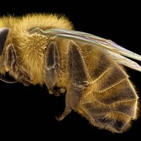
(Featured image: Honeybee, by David McCarthy and Annie Cavanagh.)

